


글로벌 연구동향
의학물리학


![[IEEE Trans Med Imaging.] Moxifloxacin-Based Extended Depth-of-Field Fluorescence Microscopy for Real-Time Conjunctival Goblet Cell Examination](/enewspaper/upimages/1665103336admin.JPG) [IEEE Trans Med Imaging.] Moxifloxacin-Based Extended Depth-of-Field Fluorescence Microscopy for Real-Time Conjunctival Goblet Cell Examination
[IEEE Trans Med Imaging.] Moxifloxacin-Based Extended Depth-of-Field Fluorescence Microscopy for Real-Time Conjunctival Goblet Cell Examination포항공대 / 이중빈, 김기현*
- 출처
- IEEE Trans Med Imaging.
- 등재일
- 2022 Aug
- 저널이슈번호
- 41(8):2004-2008. doi: 10.1109/TMI.2022.3151944.
- 내용
Abstract
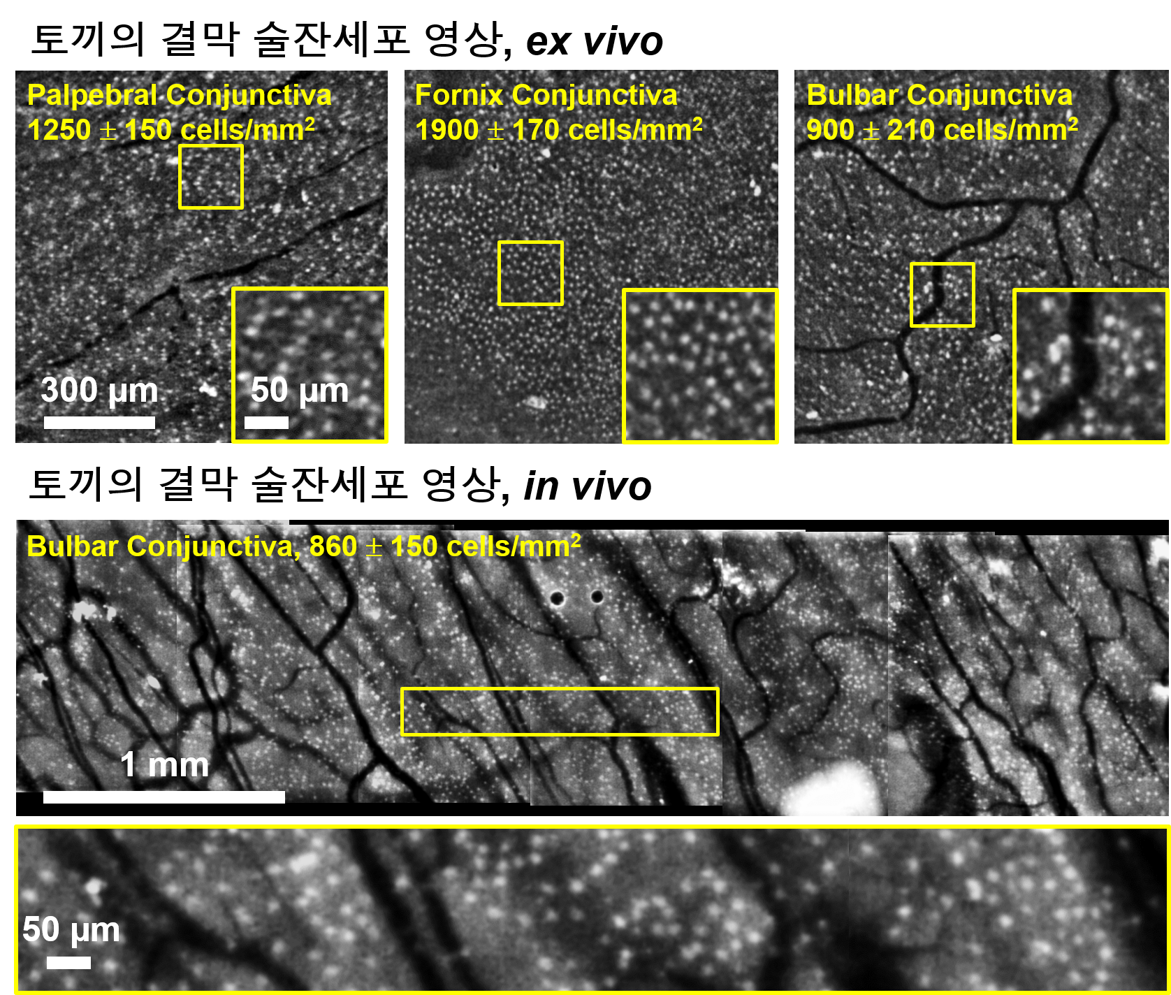
Conjunctival goblet cells (CGCs) are mucin-secreting cells in the eye and play essential roles for ocular surface homeostasis. Since various ocular surface pathologies are related to CGC dysfunction, CGC examination is important for the evaluation of ocular surface conditions. Recently we introduced moxifloxacin-based fluorescence microscopy (MBFM) for non-invasive CGC imaging. However, the imaging speed was up to 1 frame per second (fps) and needed to be improved for clinical applications. In this study, we developed a high-speed moxifloxacin-based, extended depth-of-field (EDOF) microscopy system that operates at a maximum imaging speed of 15 fps. The system used a deformable mirror for the high-speed axial sweeping of focal plane during single-frame acquisitions. The acquired images contained both in-focus and out-of-focus information, and deconvolution was used to filter the in-focus information. The system had a DOF of 800 [Formula: see text], field-of-view of 1.2 mm ×1.2 mm, and resolution of [Formula: see text]. Its performance was demonstrated by real-time, breathing-motion-insensitive CGC imaging of mouse and rabbit models, in vivo. High-speed EDOF microscopy has potentials for non-invasive, real-time CGC examinations of human subjects.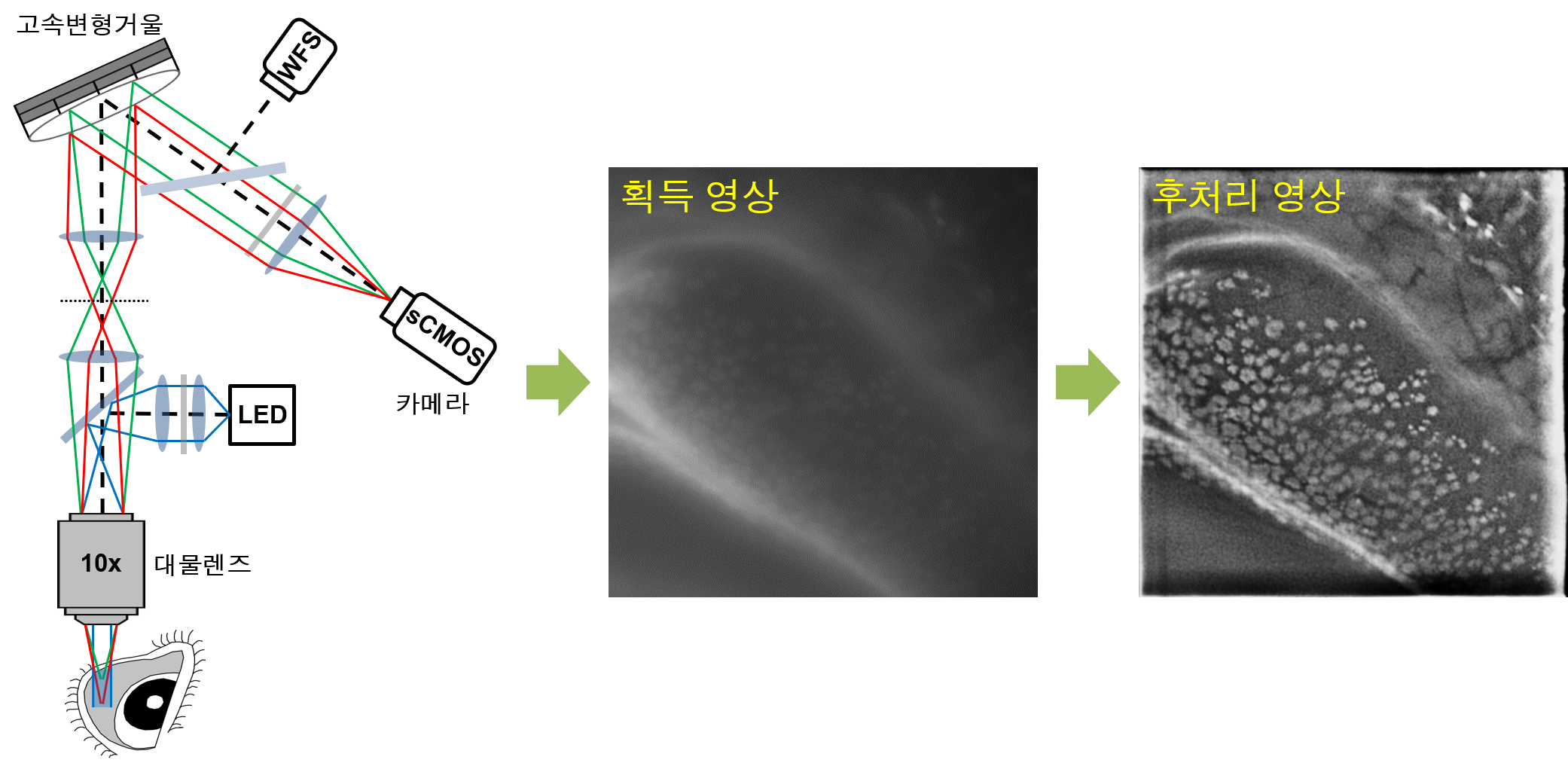
Jungbin Lee, Seonghan Kim, Jeongho Kim, Byeong Jae Son, Chang Ho Yoon, Hong Kyun Kim, Ki Hean Kim
- 연구소개
- 안구 결막 표면에 위치하며 눈물막 뮤신층 형성에 기여하는 결막 술잔세포에 대한 비침습 검사를 목표로 비접촉 실시간 영상화를 위한 현미경 영상법 개발에 관한 논문입니다. 결막 술잔세포에 대한 기존 검사법들인 침습적 압흔검사법 (impression cytology)과 비침습 공초점 반사 현미경 (cofocal reflection microscopy)은 여러 제한점 때문에 비침습 검사에 활용되지 못합니다. 본 논문에서 발표한 현미경 영상법은 기존 검사법의 한계들을 해결한 고대비도 고속 고심도 비접촉 영상화 기법이며 이를 인비보 토끼 모델에 적용하여 비침습 결막 술잔세포 검사가 가능함을 검증하였습니다. 이 현미경 영상법은 목시플록사신 안과 항생제를 활용하여 결막 술잔세포를 염색하여 형광 영상화 하며, 기울어진 결막에서도 촬영하기 위하여 초점면을 고속 이송하면서 영상화 하는 고속, 고심도 영상법을 구현하였습니다. 스펙으로는 800 마이크론의 영상심도, 초당 15 프레임 속도이었으며 토끼모델에서 실시간 결막 술잔세포 영상화가 가능하였습니다. 결막 술잔세포는 다원성 질환인 건성안의 원인 중 하나여서 원인에 따른 치료를 위해 검사가 필요합니다. 또한, 결막 술잔세포의 결핍이 직접적인 원인인 다른 안구표면질환들의 검사에 활용될 것으로 기대되어 안과 연구자들에게 도움이 될 정보라 생각합니다. 사람에게 적용 가능하며 고해상도 세포 영상화를 통한 검사법 이어서 기초 의학을 위한 고해상도 영상기기를 연구하는 연구자들에게도 도움이 될 것이라고 생각합니다.
- 덧글달기
- 이전글 [Med Phys.] Automated counting of cerebral penetrating vessels using optical coherence tomography images of a mouse brain in vivo
- 다음글 [J Appl Clin Med Phys.] Development of an anthropomorphic multimodality pelvic phantom for quantitative evaluation of a deep-learning-based synthetic computed tomography generation technique


![]()
- COPYRIGHT(C) 2015 한국원자력의학원 전략기획팀 All rights Reserved.
- 문의 : rmwebzine@kirams.re.kr 발행처 : 한국원자력의학원 전략기획팀
- 우) 01812 서울시 노원구 노원로 75 한국원자력의학원 전략기획팀