


글로벌 연구동향
의학물리학


![[Med Phys.] Automated counting of cerebral penetrating vessels using optical coherence tomography images of a mouse brain in vivo](/enewspaper/upimages/1665103591admin.JPG) [Med Phys.] Automated counting of cerebral penetrating vessels using optical coherence tomography images of a mouse brain in vivo
[Med Phys.] Automated counting of cerebral penetrating vessels using optical coherence tomography images of a mouse brain in vivo울산의대 / 최우준, 김준기*
- 출처
- Med Phys.
- 등재일
- 2022 Aug
- 저널이슈번호
- 49(8):5225-5235. doi: 10.1002/mp.15775. Epub 2022 Jun 8.
- 내용
Abstract
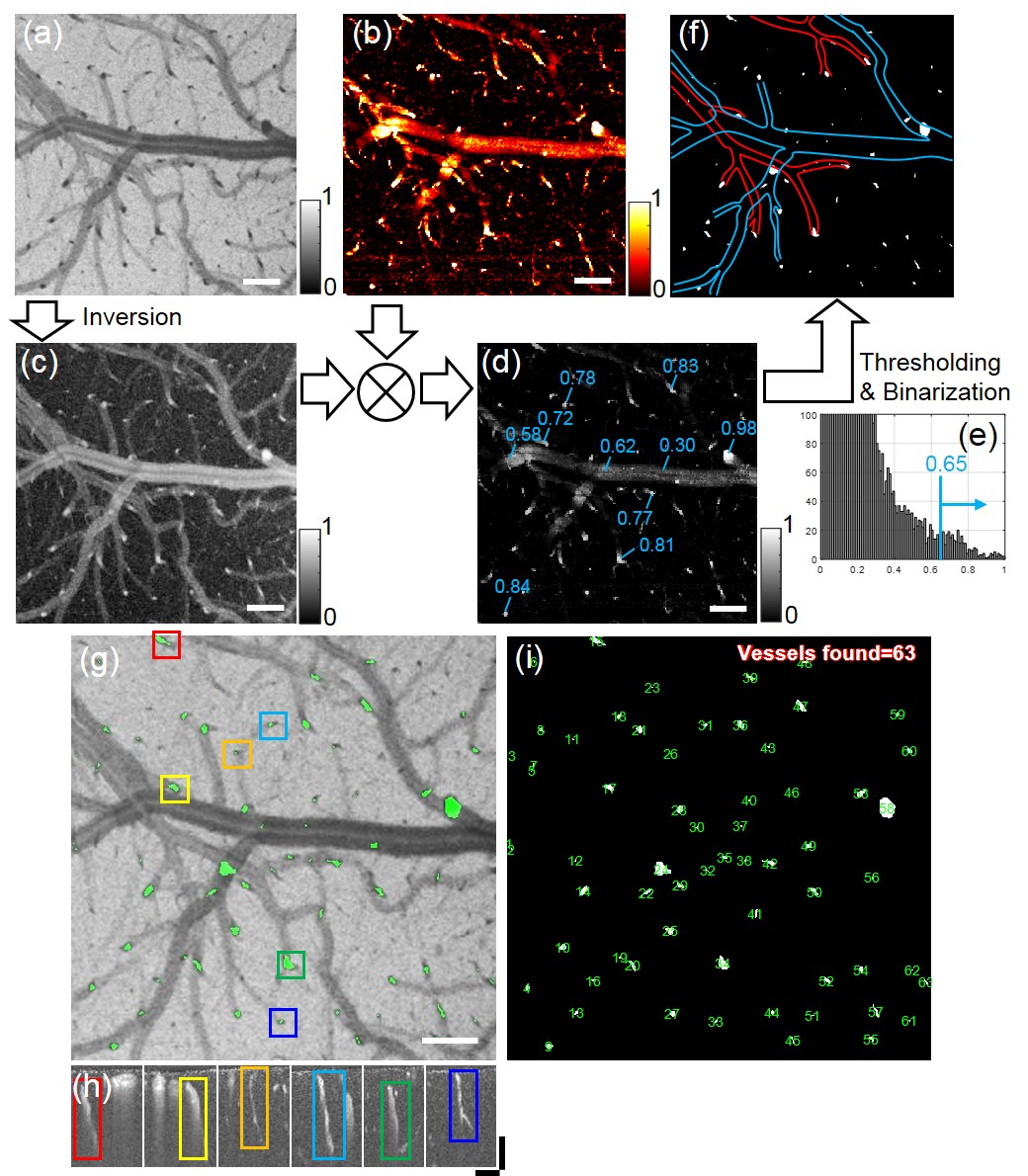
Rationale and objectives: Penetrating blood vessels emanating from cortical surface vasculature and lying deep in the cortex are essential vascular conduits for the shuttling of blood from superficial pial vessels to the capillary beds in parenchyma for the nourishment of neuronal brain tissues. Locating and counting the penetrating vessels is beneficial for the quantification of a course of ischemia in blood occlusive events such as stroke. This paper seeks to demonstrate and validate a method for automated penetrating vessel counting that uses optical coherence tomography (OCT).Materials and methods: This paper proposes an OCT method that effectively identifies and grades the cortical penetrating vessels in perfusion. The key to the proposed method is the harnessing of vascular features found in the penetrating vessels, which are distinctive from those of other vessels. In particular, with an increase in the light attenuation and flow turbulence, the contrast in the mean projection of the OCT datacube decreases, whereas that in the maximum projection of the Doppler frequency variance datacube increases. By multiplying the inversion of the former with the latter, its binary thresholding is sufficient to highlight the penetrating vessels and allows for their counting over the projection image.
Results: A computational method that leverages the decrease in mean OCT projection intensity and the increase in Doppler frequency variance at the penetrating vessel is developed. It successfully identifies and counts penetrating vessels with a high accuracy of over 87%. The penetrating vessel density is observed to be significantly reduced in the mouse model of focal ischemic stroke.
Conclusion: The OCT analysis is effective for counting penetrating blood vessels in mice brains and may be applied to the rapid diagnosis and treatment of stroke in stroke models of small animals.
Affiliations
Woo June Choi 1 , Yuandong Li 2 , Ruikang K Wang 2 , Jun Ki Kim 3 4
1 School of Electrical and Electronics Engineering, Chung-Ang University, Seoul, Republic of Korea.
2 Department of Bioengineering, University of Washington, Seattle, Washington, USA.
3 Department of Convergence Medicine, University of Ulsan College of Medicine, Seoul, Republic of Korea.
4 Asan Institute for Life Science, Asan Medical Center, Seoul, Republic of Korea.
- 키워드
- 14.11 Segmentation; 14.3 Optical computed tomography; 17.1 Image processing; 17.7 Classification methods; 17.8 Image segmentation; Doppler frequency variance; automated vessel counting; cerebral penet
- 연구소개
- 대뇌동맥에서 수직으로 분지하는 작은 관통 혈관(penetrating vessels)은 대뇌피질 내 수많은 모세혈관에 혈액을 공급해줌으로써 뇌세포 생장에 필수적인 영양분을 제공해주는 혈관입니다. 만약 이러한 관통 혈관들의 혈류 흐름이 저하되거나 폐쇄될 경우, 모세혈관에 허혈(ischemia)이 발생되고 이는 뇌조직의 괴사를 야기시켜 심각한 뇌손상을 유발할 수 있습니다. 따라서 정상 혈류를 가진 관통 혈관을 평가하는 것이 허혈성 뇌졸중을 진단하고 뇌손상 경과를 모니터링하는데 도움이 될 수 있습니다. 이에 본 논문에서는 광단층 혈관 조영술 (optical coherence tomography angiography: OCTA)을 이용하여 살아있는 소동물의 대뇌 관통 혈관들의 위치와 갯수를 자동으로 측정할 수 있는 새로운 혈관 카운팅 방법을 제안하고 있습니다. 보다 구체적으로, OCTA 데이터에서 발견되는 관통 혈관의 구조적, 유체역학적 특성에 기인한 독특한 광학 파라미터들 (높은 광감쇠와 도플러 주파수 변화)을 활용하여 수많은 뇌혈관들 중에 오로지 정상 혈류가 흐르는 관통 혈관만을 추출할 수 있습니다.
- 덧글달기


![]()
- COPYRIGHT(C) 2015 한국원자력의학원 전략기획팀 All rights Reserved.
- 문의 : rmwebzine@kirams.re.kr 발행처 : 한국원자력의학원 전략기획팀
- 우) 01812 서울시 노원구 노원로 75 한국원자력의학원 전략기획팀