


글로벌 연구동향
의학물리학


![[J Appl Clin Med Phys.] Development of an anthropomorphic multimodality pelvic phantom for quantitative evaluation of a deep-learning-based synthetic computed tomography generation technique](/enewspaper/upimages/1665103447admin.JPG) [J Appl Clin Med Phys.] Development of an anthropomorphic multimodality pelvic phantom for quantitative evaluation of a deep-learning-based synthetic computed tomography generation technique
[J Appl Clin Med Phys.] Development of an anthropomorphic multimodality pelvic phantom for quantitative evaluation of a deep-learning-based synthetic computed tomography generation technique서울대병원 / 진형민, 김정인*
- 출처
- J Appl Clin Med Phys.
- 등재일
- 2022 Aug
- 저널이슈번호
- 23(8):e13644. doi: 10.1002/acm2.13644. Epub 2022 May 17.
- 내용
Abstract
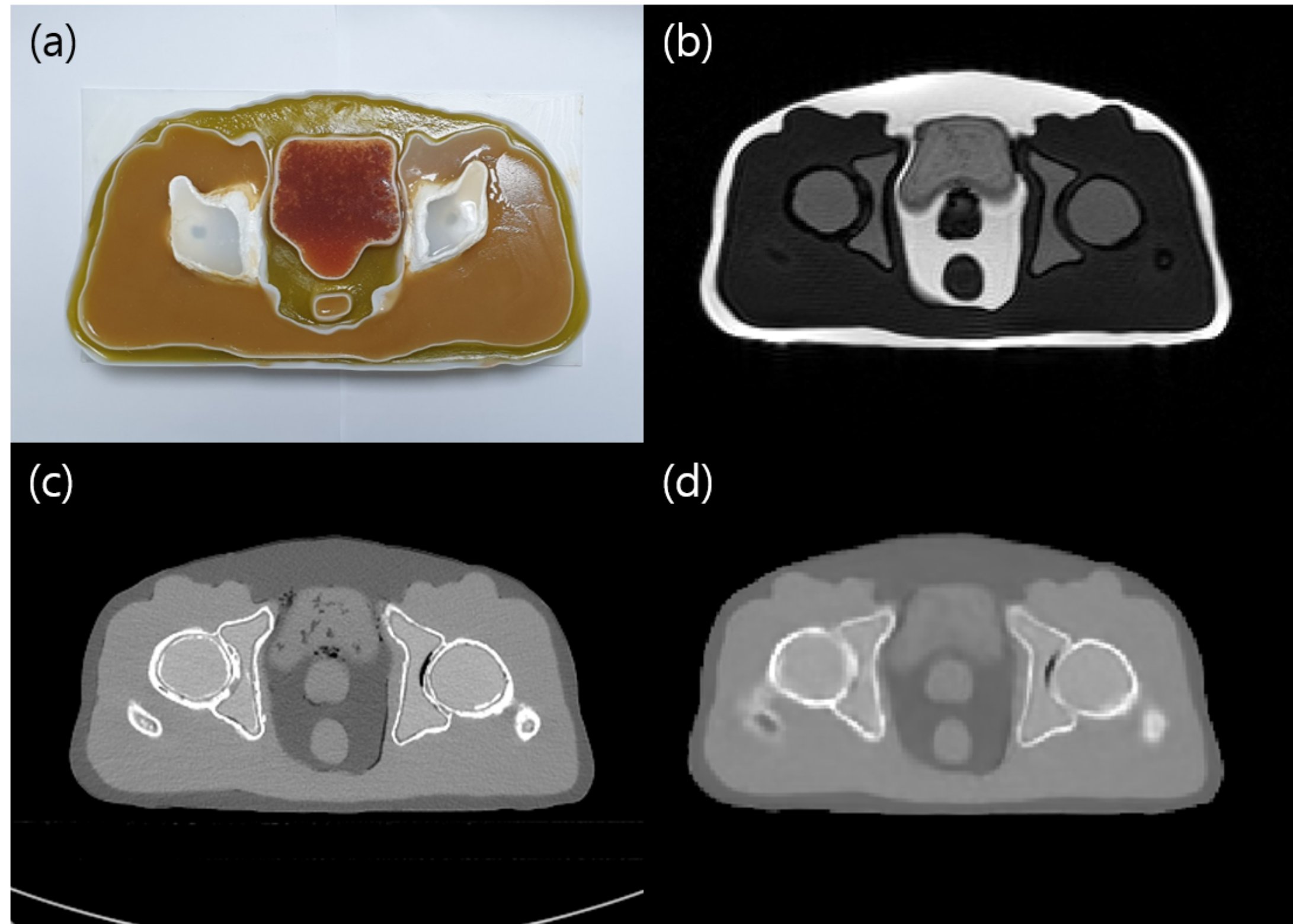
Purpose: The objective of this study was to fabricate an anthropomorphic multimodality pelvic phantom to evaluate a deep-learning-based synthetic computed tomography (CT) algorithm for magnetic resonance (MR)-only radiotherapy.Methods: Polyurethane-based and silicone-based materials with various silicone oil concentrations were scanned using 0.35 T MR and CT scanner to determine the tissue surrogate. Five tissue surrogates were determined by comparing the organ intensity with patient CT and MR images. Patient-specific organ modeling for three-dimensional printing was performed by manually delineating the structures of interest. The phantom was finally fabricated by casting materials for each structure. For the quantitative evaluation, the mean and standard deviations were measured within the regions of interest on the MR, simulation CT (CTsim ), and synthetic CT (CTsyn ) images. Intensity-modulated radiation therapy plans were generated to assess the impact of different electron density assignments on plan quality using CTsim and CTsyn . The dose calculation accuracy was investigated in terms of gamma analysis and dose-volume histogram parameters.
Results: For the prostate site, the mean MR intensities for the patient and phantom were 78.1 ± 13.8 and 86.5 ± 19.3, respectively. The mean intensity of the synthetic image was 30.9 Hounsfield unit (HU), which was comparable to that of the real CT phantom image. The original and synthetic CT intensities of the fat tissue in the phantom were -105.8 ± 4.9 HU and -107.8 ± 7.8 HU, respectively. For the target volume, the difference in D95% was 0.32 Gy using CTsyn with respect to CTsim values. The V65Gy values for the bladder in the plans using CTsim and CTsyn were 0.31% and 0.15%, respectively.
Conclusion: This work demonstrated that the anthropomorphic phantom was physiologically and geometrically similar to the patient organs and was employed to quantitatively evaluate the deep-learning-based synthetic CT algorithm.
Affiliations
Hyeongmin Jin 1 , Sung Young Lee 1 , Hyun Joon An 1 2 3 , Chang Heon Choi 1 2 3 , Eui Kyu Chie 1 2 3 4 , Hong-Gyun Wu 1 2 3 4 , Jong Min Park 1 2 3 4 5 , Sukwon Park 6 , Jung-In Kim 1 2 3
1 Department of Radiation Oncology, Seoul National University Hospital, Seoul, Republic of Korea.
2 Institute of Radiation Medicine, Seoul National University Medical Research Center, Seoul, Republic of Korea.
3 Biomedical Research Institute, Seoul National University Hospital, Seoul, Republic of Korea.
4 Department of Radiation Oncology, Seoul National University College of Medicine, Seoul, Republic of Korea.
5 Robotics Research Laboratory for Extreme Environments, Advanced Institute of Convergence Technology, Suwon, Republic of Korea.
6 Department of Radiation Oncology, Myongji Hospital, Goyang-si, Gyeonggi-do, Republic of Korea.
- 키워드
- 3D printing; deep learning; magnetic resonance-guided radiotherapy; multimodality phantom; synthetic CT.
- 연구소개
- MR 영상기반 방사선 치료계획을 세울 때 환자의 추가적인 방사선피폭을 줄이고자 딥러닝 기반의 합성 CT 생성 알고리즘이 개발되고 있습니다. 합성 CT 생성 알고리즘 평가를 위해 환자의 CT와 MR 영상을 정합하여 사용하고 있으나, 정합 알고리즘의 오차가 성능 평가에 포함되기 때문에 정형화된 팬텀을 이용한 검증이 요구되고 있습니다. 본 연구에서는 MR과 CT에서 모두 사용 가능한 골반 팬텀을 제작하여 딥러닝 기반 합성 CT 생성 알고리즘을 정량적으로 평가하고자 하였습니다. 폴리우레탄 및 실리콘 계열의 물질에 다양한 농도의 실리콘 오일을 첨가하여, CT와 MR 영상에서 인체조직과 유사한 Contrast를 얻을 수 있는 조건을 확인하였습니다. 3D printer를 이용하여 환자와 유사한 구조를 갖는 구조물에 선정된 재료를 주조하여 골반 팬텀을 제작하였습니다. 제작한 팬텀을 CT와 MR 영상을 촬영하였고, 이를 기반으로 합성 CT 영상을 생성하여 정량적으로 평가하였습니다. 장기별 CT 영상과 합성 CT 영상 간에 HU값은 유사한 수준을 보였으며, 합성 CT 영상을 이용하여 선량 계산을 하였을 때, Dose-volume histogram의 파라미터도 기존과 유사한 수준을 보였습니다. 따라서 본 연구를 통해 개발한 팬텀을 이용하여 딥러닝 기반 합성 CT 생성 알고리즘을 평가하면, 기존 평가 방법의 한계를 극복할 수 있을 것으로 생각합니다.
- 덧글달기
- 이전글 [IEEE Trans Med Imaging.] Moxifloxacin-Based Extended Depth-of-Field Fluorescence Microscopy for Real-Time Conjunctival Goblet Cell Examination
- 다음글 [Journal of the Korean Physical Society] Geant4 electromagnetic physics model assessment for RBE‑weighted dose calculation of carbon‑ion radiotherapy


![]()
- COPYRIGHT(C) 2015 한국원자력의학원 전략기획팀 All rights Reserved.
- 문의 : rmwebzine@kirams.re.kr 발행처 : 한국원자력의학원 전략기획팀
- 우) 01812 서울시 노원구 노원로 75 한국원자력의학원 전략기획팀