


글로벌 연구동향
핵의학


![[Neoplasia .] Visualizing mast cell migration to tumor sites using sodium iodide symporter of nuclear medicine reporter gene](/enewspaper/upimages/1696577305admin.JPG) [Neoplasia .] Visualizing mast cell migration to tumor sites using sodium iodide symporter of nuclear medicine reporter gene
[Neoplasia .] Visualizing mast cell migration to tumor sites using sodium iodide symporter of nuclear medicine reporter gene대구경북첨단의료산업진흥재단 / 이재언, 최준영, 전소연, 전용현*
- 출처
- Neoplasia .
- 등재일
- 2023 Sep
- 저널이슈번호
- 43:100925. doi: 10.1016/j.neo.2023.100925. Epub 2023 Aug 9.
- 내용
Abstract
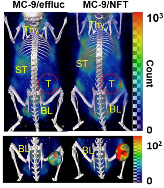
Purpose: Owing to the close relationship between mast cells and cancer progression, an imaging technique that can be applied in a clinical setting to explore the biological behavior of mast cells in the tumor microenvironment is needed. In this study, we visualized mast cell migration to lung tumor lesions in live mice using sodium iodide symporter (NIS) as a nuclear medicine reporter gene.Experimental design: The murine mast cell line MC-9 was infected with retrovirus including NIS, luciferase (as a surrogate marker for NIS), and Thy1.1 to generate MC-9/NFT cells. Radioiodine uptake was measured in MC-9/NFT cells, and an inhibition assay of radioiodine uptake using KCLO4 was also performed. Cell proliferation and FcεRI expression was examined in MC-9 and MC-9/NFT cells. The effect of mast cell-conditioned media (CM) on the proliferation of Lewis lung cancer (LLC) cells was examined. The migration level of MC-9/NFT cells was confirmed in the presence of serum-free media (SFM) and CM of cancer cells. After intravenous injection of MC-9/NFT cells into mice with an LLC tumor, I-124 PET/CT and biodistribution analysis was performed.
Results: MC-9/NFT cells exhibited higher radioiodine avidity compared to parental MC-9 cells; this increased radioiodine avidity in MC-9/NFT cells was reduced to basal level by KCLO4. Levels of FcεRI expression and cell proliferation were not different in parental MC-9 cell and MC-9/ NFT cells. The CM of MC-9/NFT cells increased cancer cell proliferation relative to that of the SFM. The migration level of MC-9/NFT cells was higher in the CM than the SFM of LLC cells. PET/CT imaging with I-124 clearly showed infiltration of reporter mast cells in lung tumor at 24 h after transfer, which was consistent with the findings of the biodistribution examination.
Conclusion: These findings suggest that the sodium iodide symporter can serve as a reliable nuclear medicine reporter gene for non-invasively imaging the biological activity of mast cells in mice with lung tumors. Visualizing mast cells in the tumor microenvironment via a nuclear medicine reporter gene would provide valuable insights into their biological functions.
I-124 PET/CT로 폐암 병변으로 비만세포의 침윤 영상화
Affiliations
Seul-Gi Oh 1, Jun Young Choi 2, Jae-Eon Lee 2, SoYeon Jeon 3, Bo-Ra Lee 3, Kwang Hee Son 3, Sang Bong Lee 4, Beum-Soo An 5, Dae Youn Hwang 5, Seong-Jang Kim 6, Ki-Tae Ha 7, Jaetae Lee 4, Yong Hyun Jeon 8
1Department of Nuclear Medicine, Kyungpook National University Hospital, Daegu, Republic of Korea; Institute of Breast Cancer Precision Medicine, Yonsei University College of Medicine, Seoul, Republic of Korea.
2Preclincial Research Center (PRC), Daegu-Gyeongbuk Medical Innovation Foundation (K-MEDI hub), Daegu, Republic of Korea; Department of Biomaterials Science (BK21 FOUR Program), Life and Industry Convergence Research Institute, College of Natural Resources and Life Science, Pusan National University, Miryang 50463, Republic of Korea.
3Preclincial Research Center (PRC), Daegu-Gyeongbuk Medical Innovation Foundation (K-MEDI hub), Daegu, Republic of Korea.
4Department of Nuclear Medicine, Kyungpook National University Hospital, Daegu, Republic of Korea.
5Department of Biomaterials Science (BK21 FOUR Program), Life and Industry Convergence Research Institute, College of Natural Resources and Life Science, Pusan National University, Miryang 50463, Republic of Korea.
6Pusan National University College of Medicine, Pusan National University School of Medicine, Busan, Republic of Korea.
7Department of Korean Medicine, School of Korean Medicine, Pusan National University, Yangsan 50612, Gyeongsangnam-do, Republic of Korea.
8Preclincial Research Center (PRC), Daegu-Gyeongbuk Medical Innovation Foundation (K-MEDI hub), Daegu, Republic of Korea. Electronic address: jeon9014@kmedihub.re.kr.
- 키워드
- Cancer; Mast cells; Nuclear reporter gene imaging; Sodium iodide symporter.
- 연구소개
- 비만세포는 점막, 피부 등 환경과 접촉하는 부위에 존재하며, 천식, 아토피 피부염 등 다양한 염증반응에 중요한 역할을 하는 면역세포이다. 비만세포는 염증반응뿐만 아니라 종양 환경에서 암 증식 촉진에도 영향을 미친다고 보고 되어 있습니다. 본 연구진은 NIS (핵의학영상 리포터 유전자)를 생체영상기술을 이용하여 종양 미세 환경에서 비만세포의 이동 및 새로운 생물학적 역할을 성공적으로 추적하였습니다. 본 연구 결과는 종양 미세 환경뿐만 아니라 알레르기 및 염증에서도 비만세포의 새로운 역할을 탐색하기 위한 새로운 접근방식을 제시한 좋은 정보라 생각합니다.
- 덧글달기


![]()
- COPYRIGHT(C) 2015 한국원자력의학원 전략기획팀 All rights Reserved.
- 문의 : rmwebzine@kirams.re.kr 발행처 : 한국원자력의학원 전략기획팀
- 우) 01812 서울시 노원구 노원로 75 한국원자력의학원 전략기획팀
편집위원
FDG NIS 리포터 유전자를 이용하여 미만세포의 이동의 영상화하는 분자영상연구로 비만세포가 종양미세환경으로서의 역할을 보여주는데 도움을 주는 연구임. 이는 핵의학 분자영상 및 종양미세환경관련 연구자들에게 관심을 끌 흥미로운 연구로 생각됨.
2023-10-06 16:16:34