


글로벌 연구동향
핵의학


- [Eur J Radiol.] Development of automated segmentation of visceral adipose tissue in computed tomography
부산대, 부산대병원 / 황재준*, 박경준*
- 출처
- Eur J Radiol.
- 등재일
- 2022 Oct 17
- 저널이슈번호
- 157:110559.
- 내용
Abstract
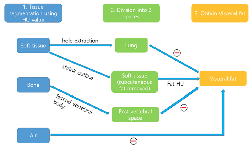
Purpose: Imaging modalities such as computed tomography (CT) or magnetic resonance imaging have been used to measure adiposity. However, manual segmentation of visceral adipose tissue (VAT) in the entire abdomen is laborious and time-consuming. We aimed to develop a new method for accurate visceral fat segmentation by automatically dividing the three anatomical compartments of the lung, soft tissue, and post-vertebral spaces.Methods: To automatically separate visceral fat, a three-step process was performed that sequentially divided tissues and regions in a three-dimensional CT image. Manual segmentation was performed in 99 individuals who underwent 18-fluoro-2-deoxyglucosepositron emission tomography/CT for cancer screening between January 2010 and December 2018 to validate the automated segmentation. The similarity index and Pearson's correlation analysis were performed to compare automated segmentation with manual segmentation. Clinical data, such as weight, height, and glucose and insulin levels, were measured. Pearson's correlation analysis was performed to investigate the association between the two methods.
Results: VAT volume of automated segmentation (3,594.6 ± 1,776.5 cm3) strongly correlated with that of manual segmentation (3,375.7 ± 1567.5 cm3) (r = 0.9676, p < 0.0001). The similarity index positively correlated with the VAT volume (r = 0.6396, p < 0.0001) and negatively correlated with the mean Hounsfield units (HU) (r = -0.4328, p < 0.0001). Bland-Altman plots are presented with 5.1 % for VAT volume and 7.1 % for mean HU were outside 1.96 standard deviation from the mean value.
Conclusion: We developed an automated segmentation method for VAT in the entire abdomen. This automated segmentation method is feasible for measuring the VAT volume and VAT HU. This method could be employed in daily clinical practice to provide more detailed information about VAT.
Affiliations
Jae Joon Hwang 1, Kyoungjune Pak 2
1Department of Oral and Maxillofacial Radiology and Dental Research Institute, Pusan National University, Yangsan, Republic of Korea; Dental and Life Science Institute & Dental Research Institute, School of Dentistry, Pusan National University, Yangsan, Republic of Korea. Electronic address: softdent@pusan.ac.kr.
2Department of Nuclear Medicine and Biomedical Research Institute, Pusan National University Hospital, Busan, Republic of Korea. Electronic address: ilikechopin@pusan.ac.kr.
- 키워드
- Abdominal fat; Computed tomography; Obesity; Software.
- 연구소개
- 검진으로 시행된 F18-FDG PET/CT의 CT 영상에서 내장지방의 부피를 측정하는 방법을 새롭게 개발한 연구입니다. 대부분의 기존 연구들이 내장지방의 단면 표면적을 측정하였으나, 이 연구에서는 automated segmentation을 통한 부피 측정으로, 더욱 정확히 내장지방에 대한 정보를 얻을 수 있습니다. 총 99명의 CT 영상에서, 새롭게 개발된 automated segmentation 방법을 manual segmentation 방법과 비교하여, 정확성 및 측정 시간 단축(약 30초)을 확인할 수 있었습니다.
- 덧글달기
- 이전글 [Sci Rep.] Reliability and feasibility of visual grading systems and quantitative indexes on [99mTc]Tc-DPD imaging for cardiac amyloidosis
- 다음글 [Medicina (Kaunas) .] Hepatic Fat Quantification with the Multi-Material Decomposition Algorithm by Using Low-Dose Non-Contrast Material-Enhanced Dual-Energy Computed Tomography in a Prospectively Enrolled Cohort


![]()
- COPYRIGHT(C) 2015 한국원자력의학원 전략기획팀 All rights Reserved.
- 문의 : rmwebzine@kirams.re.kr 발행처 : 한국원자력의학원 전략기획팀
- 우) 01812 서울시 노원구 노원로 75 한국원자력의학원 전략기획팀
편집위원
최근 대사질환 및 종양미세환경 관련 지방세포의 분석이 활발히 이루어지고 있는데 임상에서 간편하게 수행할 수 있으며 정확도도 높은 VAT 측정법이 관련 연구 활성화에 도움이 될 것으로 기대됨.
2023-02-06 15:50:50