
글로벌 연구동향
핵의학


![[Exp Cell Res.] Role of M2-like macrophages in the progression of ovarian cancer](/enewspaper/upimages/1606889649admin.JPG) 2020년 12월호
2020년 12월호
[Exp Cell Res.] Role of M2-like macrophages in the progression of ovarian cancer경북의대 / 백세환, 안병철*
- 출처
- Exp Cell Res.
- 등재일
- 2020 Oct 15
- 저널이슈번호
- 395(2):112211. doi: 10.1016/j.yexcr.2020.112211. Epub 2020 Aug 2.
- 내용
Abstract
In this study, we noninvasively assessed whether M2-like macrophages accelerate the progression of ovarian cancer by performing molecular imaging of ovarian cancer cells expressing enhanced firefly luciferase (Effluc) in living mice. First, murine ovarian cancer ID8 cells expressing Effluc (ID8/Effluc cells) were established by retroviral infection. Subsequently, macrophages were isolated from the peritoneal exudate of mice injected with thioglycollate medium and differentiated into M2-like macrophages by adding interleukin 4. To characterize these M2-like macrophages, F4/80 and cluster of differentiation 206 expression levels were determined. Then, the M2-like macrophages were co-cultured with the ID8/Effluc cells and bioluminescence imaging (BLI) of signals from the ID8/Effluc cells was completed. Additionally, migration and wound healing were assessed to evaluate the effects of conditioned medium (CM) from M2-like macrophages on ID8/Effluc cell motility. In the in vivo study, mice were first given either liposome-phosphate-buffered saline or liposome-clodronate (lipo-clodronate). After 24 h, ID8/Effluc cells were intraperitoneally injected into the mice and BLI was completed at the designed time points. Next, histological analysis was conducted to characterize the infiltrated tumor. Flow cytometric analysis revealed high levels of CD206 expression in the differentiated M2-like macrophages. Meanwhile, ID8/Effluc cells co-cultured with these M2-like macrophages proliferated rapidly in an M2-like macrophage, number-dependent manner. The migration of the ID8/Effluc cells was also increased by the application of CM from M2-like macrophages. In vivo BLI revealed that the growth rate of intraperitoneally injected ovarian cancer cells was inhibited following macrophage depletion by treatment with lipo-clodronate. M2-like macrophages accelerated the progression of ovarian cancer, suggesting they are a new therapeutic target for ovarian cancer and that ovarian cancer could be managed by altering the nature of communication between ovarian cancer and macrophages.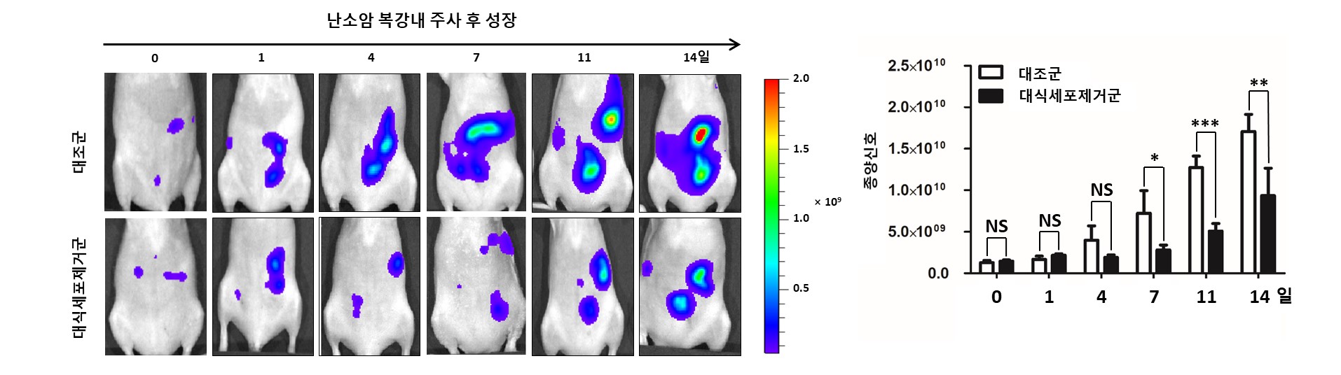
Affiliations
Se Hwan Baek 1 , Ho Won Lee 1 , Prakash Gangadaran 2 , Ji Min Oh 1 , Liya Zhu 1 , Ramya Lakshmi Rajendran 1 , Jaetae Lee 3 , Byeong-Cheol Ahn 4
1 Department of Nuclear Medicine, School of Medicine, Kyungpook National University, Daegu, Republic of Korea.
2 Department of Nuclear Medicine, School of Medicine, Kyungpook National University, Daegu, Republic of Korea; BK21 Plus KNU Biomedical Convergence Program, Department of Biomedical Science, School of Medicine, Kyungpook National University, Daegu, Republic of Korea.
3 Department of Nuclear Medicine, School of Medicine, Kyungpook National University, Daegu, Republic of Korea; Department of Nuclear Medicine, Kyungpook National University Hospital, Daegu, Republic of Korea.
4 Department of Nuclear Medicine, School of Medicine, Kyungpook National University, Daegu, Republic of Korea; BK21 Plus KNU Biomedical Convergence Program, Department of Biomedical Science, School of Medicine, Kyungpook National University, Daegu, Republic of Korea; Department of Nuclear Medicine, Kyungpook National University Hospital, Daegu, Republic of Korea. Electronic address: abc2000@knu.ac.kr.
- 연구소개
- 대식세포가 악성종양의 진행을 촉진 시키거나 억제할 수 있음에 대한 연구가 진행되고 있습니다. 해당 연구는 type 2 대식세포의 사멸시키면, 난소암의 진행을 억제할 수 있음을 동물실험을 통해 확인한 연구입니다. 종양 치료법 개발 관련 기초 및 임상 연구자에게 도움이 될 정보로 생각합니다.
- 덧글달기


편집위원
대식세포가 악성종양의 진행에 영향을 주는 것이 예상되고 있는데, 해당 연구는 대식세포의 사멸이 난소암의 진행을 억제함을 있음을 보여준 연구임. 종양관련 연구자 및 임상가에게 관심을 유도할 연구로 생각됨.
덧글달기닫기2020-12-02 11:18:05
등록