


글로벌 연구동향
핵의학


![[J Nucl Med.] N-(2-(Dimethylamino)Ethyl)-4-18F-Fluorobenzamide: A Novel Molecular Probe for High-Contrast PET Imaging of Malignant Melanoma.](/enewspaper/upimages/1569894467admin.JPG) [J Nucl Med.] N-(2-(Dimethylamino)Ethyl)-4-18F-Fluorobenzamide: A Novel Molecular Probe for High-Contrast PET Imaging of Malignant Melanoma.악성 흑색종 진단을 위한 새로운 PET 방사성의약품의 개발
[J Nucl Med.] N-(2-(Dimethylamino)Ethyl)-4-18F-Fluorobenzamide: A Novel Molecular Probe for High-Contrast PET Imaging of Malignant Melanoma.악성 흑색종 진단을 위한 새로운 PET 방사성의약품의 개발전남의대 / 표아영, 김동연*, 민정준*
- 출처
- J Nucl Med.
- 등재일
- 2019 Jul
- 저널이슈번호
- 60(7):924-929. doi: 10.2967/jnumed.118.221416. Epub 2018 Dec 14.
- 내용
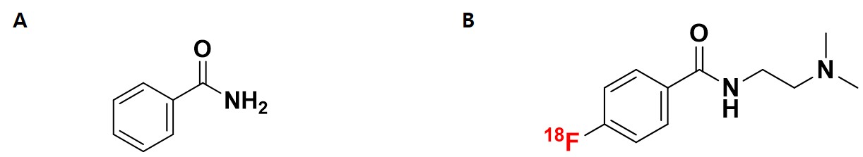
그림 1. (A) 벤즈아마이드의 화학적 구조 및 (B) N-(2-(dimethylamino)ethyl)-4-18F-fluorobenzamide ([18F]DMFB)의 화학적 구조
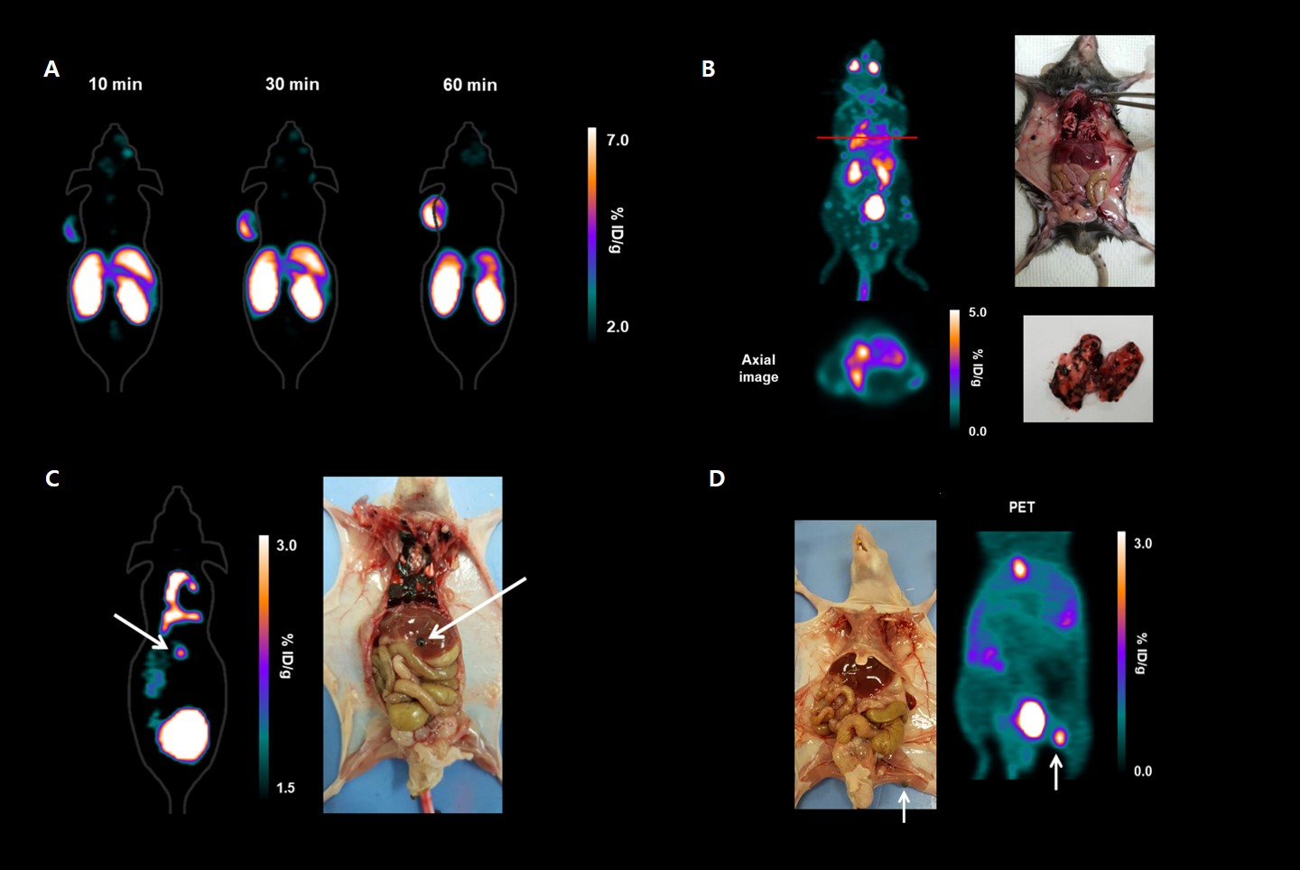
그림 2. 원발성 및 전이성 흑색종 소동물 모델을 이용한 [18F]DMFB의 영상평가. (A) 피하이식 흑색종 소동물 모델에서 [18F]DMFB 정맥주사 후 10, 30, 60분 후의 PET 영상. (B) 폐 전이, (C) 간 전이, (D) 림프절 전이 흑색종 소동물 모델에서 [18F]DMFB 정맥주사 후 60분 후의 PET 영상
Abstract
Malignant melanoma is an aggressive and serious form of skin cancer, with prognosis and treatment outcome depending heavily on the clinical stage of the disease at the time of diagnosis. Here, we synthesized a novel 18F-labeled benzamide derivative to target melanoma and then evaluated its biologic characteristics in small-animal models. Methods: N-(2-(dimethylamino)ethyl)-4-18F-fluorobenzamide (18F-DMFB) was synthesized by reaction of N-succinimidyl 4-18F-fluorobenzoate with N,N-dimethylethylenediamine. The binding affinity of 18F-DMFB was measured in B16F10 (mouse melanoma) cells with or without l-tyrosine. Small-animal PET imaging with 18F-DMFB was performed on B16F10 xenograft and metastasis mouse models. Results: The overall non-decay-corrected radiochemical yield of 18F-DMFB was approximately 10%-15%. Uptake of 18F-DMFB was melanin-specific, as cellular uptake in B16F10 increased more than 18-fold in the presence of l-tyrosine. Biodistribution studies revealed that 18F-DMFB accumulated, and was retained, in B16F10 xenografts for 120 min (10, 30, 60, and 120 min: 9.24, 10.80, 13.0, and 10.59 percentage injected dose/g, respectively) after radiotracer injection. Liver uptake of 18F-DMFB decreased from 10 to 120 min and showed fast clearance (10, 30, 60, and 120 min: 11.19, 5.7, 2.47, and 0.4 percentage injected dose/g). Furthermore, 18F-DMFB allowed visualization of metastatic lesions immediately after injection and was retained in lesions for over 60 min, with a high tumor-to-background ratio. Conclusion: 18F-DMFB demonstrated a high melanin-targeting ability and tumor-specific tumor uptake in both primary and metastatic lesions in animal models bearing malignant melanoma. 18F-DMFB may be a potential PET imaging agent for melanoma.
Author informationPyo A1, Kim HS1, Kim HS2, Yun M3, Kim DY4, Min JJ4.
1
Department of Nuclear Medicine, Chonnam National University Medical School and Hwasun Hospital, Hwasun, Korea.
2
Department of Forensic Medicine, Chonnam National University Medical School, Hwasun, Korea; and.
3
Microbiology and Functionality Research Group, Research and Development Division, World Institute of Kimchi, Gwangju, Korea.
4
Department of Nuclear Medicine, Chonnam National University Medical School and Hwasun Hospital, Hwasun, Korea jjmin@jnu.ac.kr blueburr@gmail.com.
- 키워드
- 18F-labeled benzamide derivative; PET; malignant melanoma; metastasis; molecular imaging
- 연구소개
- 본 논문은 전이가 되면 1년내 사망률이 75%에 이를 만큼 공격적인 악성흑색종을 세포수준에서 조기진단 할 수 있는 새로운 방사성의약품의 개발에 관한 연구입니다. 저희 연구진은 악성흑색종의 중요한 바이오마커인 멜라닌을 선택적 표적 할 수 있는 벤즈아마이드 구조를 변경하여 PET 용 방사성핵종인 18F이 표지된 새로운 화합물 N-(2-(dimethylamino)ethyl)-4-18F-fluorobenzamide ([18F]DMFB)을 개발하였으며(그림 1), 이의 활성을 세포 및 여러 종류의 흑색종 소동물 모델에서 평가하여 진단제제로써의 가능성을 검증하였습니다. [18F]DMFB의 가장 큰 특징인 멜라닌에 대한 높은 선택적 섭취 및 다른 장기에서의 빠른 배출은 PET 영상에서 흑색종에 대한 매우 우수한 영상 대비효과를 줄 수 있었으며(그림 2), 이러한 점은 [18F]DMFB가 주사 후 1시간 이내에 원발성 및 전이성 흑색종을 기존의 방법보다 훨씬 정확하게 진단 할 수 있음을 의미한다고 생각합니다.
- 덧글달기
- 이전글 [Eur J Nucl Med Mol Imaging.] Prognostic value of metabolic tumour volume on baseline 18F-FDG PET/CT in addition to NCCN-IPI in patients with diffuse large B-cell lymphoma: further stratification of the group with a high-risk NCCN-IPI.
- 다음글 [PLoS One.] FDG PET/CT for the early prediction of RAI therapy response in patients with metastatic differentiated thyroid carcinoma.


![]()
- COPYRIGHT(C) 2015 한국원자력의학원 전략기획팀 All rights Reserved.
- 문의 : rmwebzine@kirams.re.kr 발행처 : 한국원자력의학원 전략기획팀
- 우) 01812 서울시 노원구 노원로 75 한국원자력의학원 전략기획팀
편집위원
N-(2-(dimethylamino)ethyl)-4-18F-fluorobenzamide (18F-DMFB)을 이용하여 동물모델에서 악성흑생색종을 잘 보여줄 수 있음을 보연준 기초 연구임. 핵의학 및 피부암 관련 임상의사 및 연구자들에게 유용한 정보를 제공하고 관심을 끌 수 있을 것으로 생각됨. 또한 종양특이섭취 정도에 따라 theranostic approach도 가능할 수 있을 것을 보이며, 이는 향후 핵의학 발전에 큰 기여를 할 수 있을 것임.
2019-08-30 10:02:52