


글로벌 연구동향
핵의학


![[Cancer Imaging.] Improved detection of metastatic lymph nodes in oesophageal squamous cell carcinoma by combined interpretation of fluorine-18-fluorodeoxyglucose positron-emission tomography/computed tomography.](/enewspaper/upimages/1564467647admin.JPG) [Cancer Imaging.] Improved detection of metastatic lymph nodes in oesophageal squamous cell carcinoma by combined interpretation of fluorine-18-fluorodeoxyglucose positron-emission tomography/computed tomography.
[Cancer Imaging.] Improved detection of metastatic lymph nodes in oesophageal squamous cell carcinoma by combined interpretation of fluorine-18-fluorodeoxyglucose positron-emission tomography/computed tomography.성균관의대 / 이지영, 김영환, 최준영*
- 출처
- Cancer Imaging.
- 등재일
- 2019 Jun 21
- 저널이슈번호
- 19(1):40. doi: 10.1186/s40644-019-0225-5.
- 내용
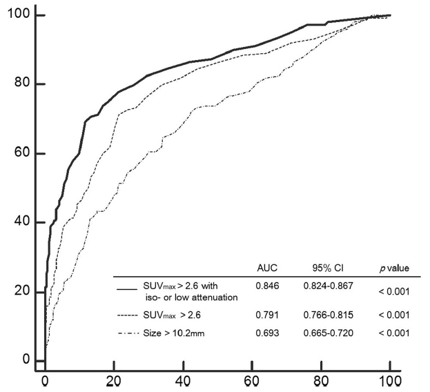
그림 설명: 림프절의 크기나 SUVmax로 진단하는 것 보나다 림프절의 SUVmax와 CT attenuation 같이 고려하는 것이 전이림프절 전이 진단능이 유의하게 향상됨.
Abstract
BACKGROUND:
We sought to evaluate the diagnostic performance of fluorine-18-fluorodeoxyglucose positron-emission tomography/computed tomography (18F-FDG PET/CT) in the detection of metastatic lymph nodes by combined interpretation of PET/CT images in patients with oesophageal squamous cell carcinoma.METHODS:
Two hundred three patients with oesophageal squamous cell carcinoma underwent 18F-FDG PET/CT before oesophagectomy and lymph node dissection. Maximum standardized uptake value (SUVmax), mean Hounsfield unit (HU), short axis diameter (size), and visual CT attenuation (high, iso-, low) were evaluated on noncontrast CT and PET images following PET/CT scan. In this combined interpretation protocol, the high attenuated lymph nodes were considered benign, even if the SUVmax value was high. The diagnostic accuracy of each method was compared using the postoperative histologic result as a reference standard.RESULTS:
A total of 1099 nodal stations were dissected and 949 nodal stations were proven to demonstrate metastasis. SUVmax and size of the malignant lymph nodes were higher than those of the benign nodes, and visual CT attenuation was significantly different among the two groups (P < 0.001). Using cutoff values of 2.6 for SUVmax and 10.2 mm for size, the combined interpretation of an SUVmax of more than 2.6 with iso- or low CT attenuation [area under the curve (AUC): 0.846, 95% confidence interval (CI): 0.824-0.867] showed significantly better diagnostic performance for detecting malignant lymph nodes than SUVmax only (AUC: 0.791, 95% CI: 0.766-0.815) and size (AUC: 0.693, 95% CI: 0.665-0.720) methods (P < 0.001) in a receiver operating characteristic curve analysis.CONCLUSIONS:
The diagnostic accuracy of PET/CT for nodal metastasis in oesophageal squamous cell carcinoma was improved by the combined interpretation of 18F-FDG uptake and visual CT attenuation pattern.
Author informationLee JY1, Kim YH2, Park YJ3, Park SB4, Chung HW5, Zo JI6, Shim YM6, Lee KS7, Choi JY8.
1
Department of Nuclear Medicine, Jeju National University Hospital, Jeju National University School of Medicine, Jeju, Republic of Korea.
2
Department of Nuclear Medicine, Kangbuk Samsung Hospital, Sungkyunkwan University School of Medicine, Seoul, Republic of Korea.
3
Department of Nuclear Medicine, Samsung Medical Center, Sungkyunkwan University School of Medicine, 81 Irwon-ro, Gangnam-gu, Seoul, 06351, Republic of Korea.
4
Department of Radiology, Soonchunhyang University Seoul Hospital, Soonchunhyang University College of Medicine, Seoul, Republic of Korea.
5
Department of Nuclear Medicine, Konkuk University Medical Center, Konkuk University School of Medicine, Seoul, Republic of Korea.
6
Department of Thoracic and Cardiovascular Surgery, Samsung Medical Center, Sungkyunkwan University School of Medicine, Seoul, Republic of Korea.
7
Department of Radiology, Samsung Medical Center, Sungkyunkwan University School of Medicine, Seoul, Republic of Korea.
8
Department of Nuclear Medicine, Samsung Medical Center, Sungkyunkwan University School of Medicine, 81 Irwon-ro, Gangnam-gu, Seoul, 06351, Republic of Korea. jynm.choi@samsung.com.
- 키워드
- 18F-FDG; CT attenuation; Lymph node metastasis; Oesophageal cancer; PET/CT; SUVmax
- 연구소개
- 식도암 초기 병기 결정 목적으로 시행한 FDG PET/CT의 전이 림프절 진단에서 림프절의 CT attenuation을 고려하여 전이여부를 진단하는 것이 PET/CT의 전이 림프절 진단능을 유의하게 향상시키는 것을 보여준 연구입니다. 우리나라 같이 결핵 등 염증폐질환의 빈도가 높은 나라에서 식도암 전이 림프절 진단에서 PET/CT의 위양성을 줄일 수 있어 임상적으로 중요한 연구입니다.
- 덧글달기


![]()
- COPYRIGHT(C) 2015 한국원자력의학원 전략기획팀 All rights Reserved.
- 문의 : rmwebzine@kirams.re.kr 발행처 : 한국원자력의학원 전략기획팀
- 우) 01812 서울시 노원구 노원로 75 한국원자력의학원 전략기획팀
편집위원
식도암의 병기설정을 위한 PET/CT판독에 대해 보다 더 정량적인 기준을 바탕으로 정확한 해석에 도움을 줄 수 있는 연구입니다.
2019-07-18 15:23:32