글로벌 연구동향
핵의학
 Adenovirus-mediated expression of human sodium-iodide symporter gene permits in vivo tracking of adipose tissue-derived stem cells in a canine myocardial infarction model.
(KIRAMS,건국대/이아라,이용진*,엄기동*)
- 출처
- Nucl Med Biol.
- 등재일
- 2015 Jul
- 저널이슈번호
- 42(7):621-9.
- 내용

[Abstract]INTRODUCTION:
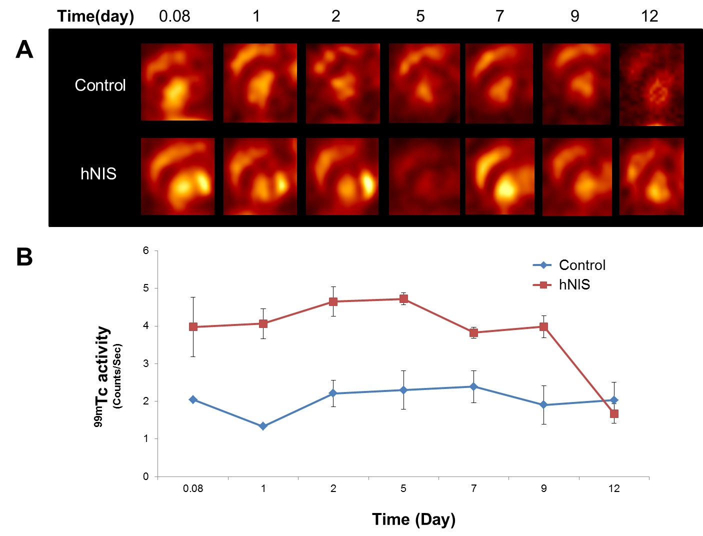
In vivo tracking of the transplanted stem cells is important in preclinical research of stem cell therapy for myocardial infarction. We examined the feasibility of adenovirus -mediated sodium iodide symporter (NIS) gene to cell tracking imaging of transplanted stem cells in a canine infarcted myocardium by clinical single photon emission computed tomography (SPECT).METHODS: Beagle dogs were injected intramyocardially with NIS-expressing adenovirus-transfected canine stem cells (Ad-hNIS-canine ADSCs) a week after myocardial infarction (MI) development. (99m)Tc-methoxyisobutylisonitrile ((99m)Tc-MIBI) and (99m)Tc-pertechnetate ((99m)TcO4(-)) SPECT imaging were performed for assessment of infarcted myocardium and viable stem cell tracking. Transthoracic echocardiography was performed to monitor any functional cardiac changes.RESULTS: Left ventricular ejection fraction (LVEF) was decreased after LAD ligation. There was no significant difference in EF between the groups with the stem cell or saline injection. (125)I uptake was higher in Ad-hNIS-canine ADSCs than in non-transfected ADSCs. Cell proliferation and differentiation were not affected by hNIS-carrying adenovirus transfection. (99m)Tc-MIBI myocardial SPECT imaging showed decreased radiotracer uptake in the infarcted apex and mid-anterolateral regions. Ad-hNIS-canine ADSCs were identified as a region of focally increased (99m)TcO4(-) uptake at the lateral wall and around the apex of the left ventricle, peaked at 2 days and was observed until day 9.CONCLUSIONS: Combination of adenovirus-mediated NIS gene transfection and clinical nuclear imaging modalities enables to trace the fate of transplanted stem cells in infarcted myocardium for translational in vivo cell tracking study for prolonged duration
[Author Information]
Lee AR1, Woo SK2, Kang SK3, Lee SY4, Lee MY4, Park NW4, Song SH4, Lee SY4, Nahm SS5, Yu JE5, Kim MH2, Yoo RJ2, Kang JH2, Lee YJ6, Eom KD7.
1Department of Veterinary Radiology and Diagnostic Imaging, College of Veterinary Medicine, Konkuk University, Seoul, Korea. Electronic address: alvetrad09@gmail.com.
2Molecular Imaging Research Center, Korea Institute of Radiological and Medical Sciences, Seoul, Korea.
3Biostar Stem Cell Research Institute, K-STEMCELL Co., Ltd, Seoul, Korea.
4Department of Veterinary Radiology and Diagnostic Imaging, College of Veterinary Medicine, Konkuk University, Seoul, Korea.
5Laboratory of Veterinary Anatomy, College of Veterinary Medicine, Konkuk University, Seoul, Korea.
6Molecular Imaging Research Center, Korea Institute of Radiological and Medical Sciences, Seoul, Korea. Electronic address: yjlee@kirams.re.kr.
7Department of Veterinary Radiology and Diagnostic Imaging, College of Veterinary Medicine, Konkuk University, Seoul, Korea. Electronic address: eomkd@konkuk.ac.kr
- 연구소개
- 줄기세포를 이용한 난치성 질환 치료 연구와 관련하여 이식한 줄기세포를 추적하고자 하는 영상기법이 활발히 진행되고 있다. 그러나 이식된 줄기세포의 생존, 이동 및 분화에 관한 생체내 추적연구는 상대적으로 미흡한 실정이다. 본 연구는 치료용 줄기세포에 핵의학 영상 구현을 위한 리포터 유전자를 이입 후 심근경색질환 모델 비글견 심근에 직접 투여 후 동위원소를 이용한 핵의학적 방법(SPECT)으로 세포 추적을 한 결과, 반복적으로 영상화 재현이 가능하다는 결론을 얻을 수 있었다. 향후 개발되고 있는 치료용 줄기세포의 성능 및 생체 내 평가를 함에 있어 임상 및 비임상에 적용하다고 판단되며, 치료용 줄기세포 개발에 가속화 시킬 수 있는 토대가 될 것으로 기대된다.
- 덧글달기


